- Differential Interference Contrast
-
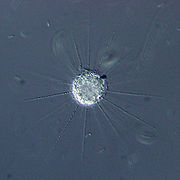 DIC-Aufnahme des Sonnentierchens Raphidiophrys contractilis
Der Differentialinterferenzkontrast (auch: Differential-Interferenz-Kontrast oder Nomarski-Kontrast, abgekürzt DIC von englisch Differential Interference Contrast) ist eine Methode der abbildenden Durchlicht-Mikroskopie, die Unterschiede in der optischen Dichte des betrachteten Objektes in Kontrastunterschiede des Bildes umwandelt. Mit diesem Verfahren lassen sich sehr eindrucksvolle pseudo-plastische Bilder erstellen, die allerdings nicht die wahren räumlichen Strukturen wiedergeben.
Inhaltsverzeichnis
Geschichte
DIC wurde in den 1950er Jahren von Georges Nomarski in Paris entwickelt; das CNRS hielt die Lizenz für dieses Verfahren. Die erste serienmäßige Anwendung baute die Firma Carl Zeiss in Oberkochen. In der Folgezeit haben nur die großen Mikroskophersteller das anspruchsvolle Verfahren in ihr Programm übernommen.
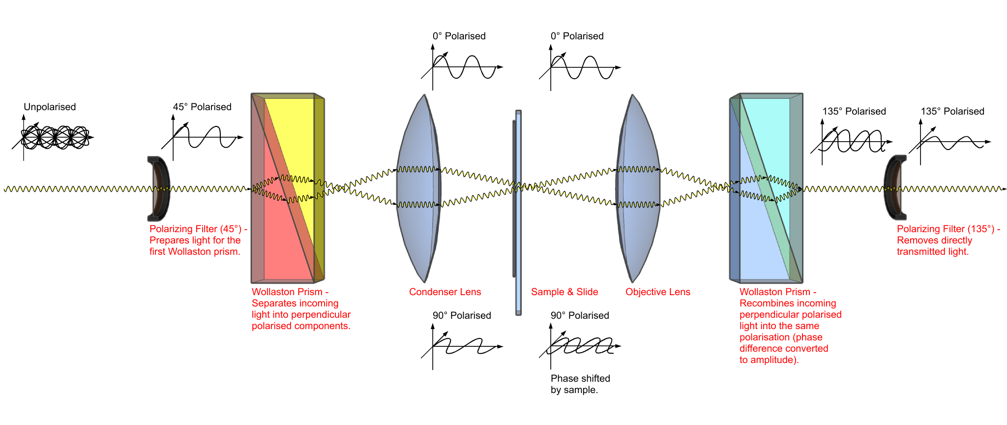
Prinzip
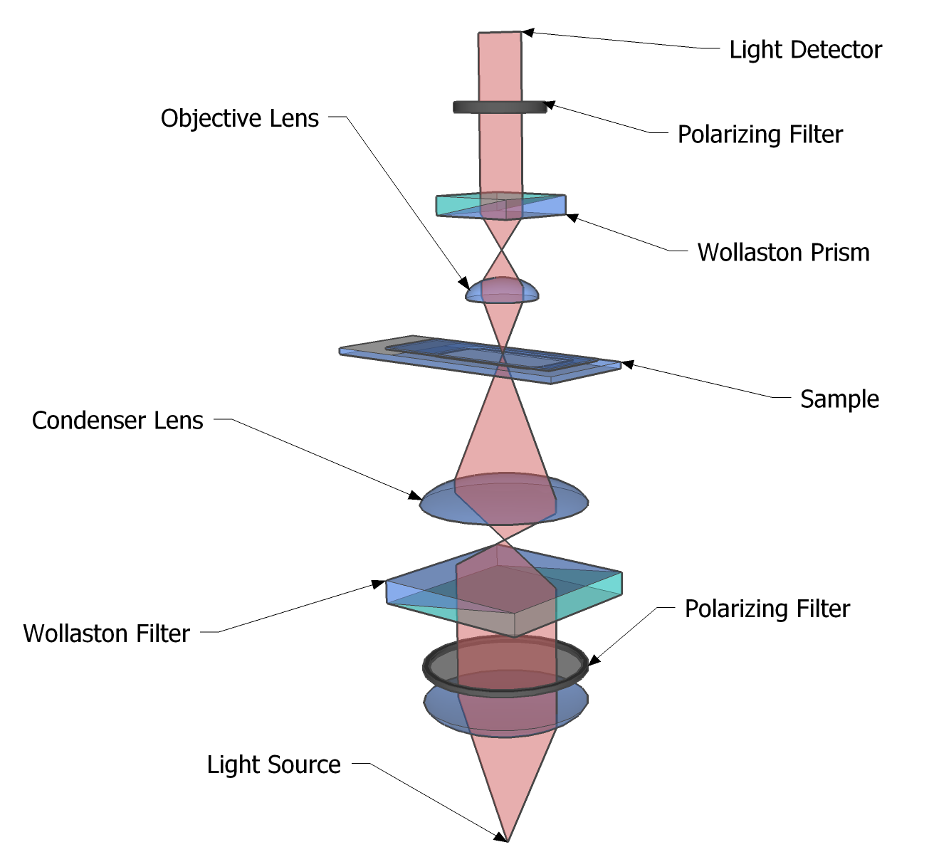
 Mikroskopische Komponenten bei DIC-Mikroskopie. Statt eines Wollaston-Prismas wird in der Regel ein Nomarski-Prisma verwendet.
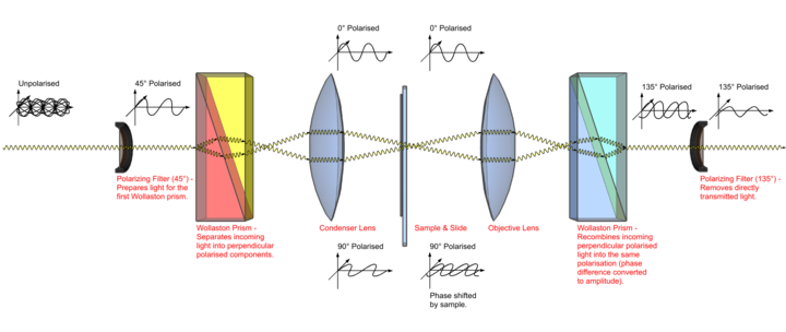
Neben dem üblichen Mikroskopaufbau werden zusätzlich je ein Nomarski-Prisma, das aus zwei Kalkspatkeilen besteht, und je ein Polarisationsfilter vor dem Kondensor und hinter dem Objektiv eingebaut. Das Kondensor-Prisma sorgt für die Aufspaltung des Beleuchtungsstrahlbündels in zwei parallele, senkrecht zueinander schwingende Strahlen, die einen Versatz unterhalb der Auflösungsgrenze des Mikroskopobjektives aufweisen. Beide Strahlen werden nach dem Durchgang durch Präparat und Objektiv im darüber befindlichen Objektiv-Prisma wieder zusammengeführt und können so interferieren, nachdem ihre Polarisationsrichtungen durch den Filter vereinigt wurden. Falls die beiden Teilstrahlen durch ein Gebiet unterschiedlicher optischer Dichte gelaufen waren, wird ein Kontrast erzeugt (destruktive Interferenz, ansonsten wird die ursprüngliche Intensität vor der Strahlteilung wieder erreicht.
Da die Teilstrahlen senkrecht zueinander polarisiert sind, werden durch Drehung des Mikroskoptisches unterschiedliche Darstellungen des Objektes möglich. Durch Einbau eines λ/4-Plättchens kann zusätzlich ein Farbkontrast erzeugt werden.
Im Gegensatz zur Phasenkontrastmikroskopie kann die maximale Kondensor-Apertur verwendet werden. Dadurch steigt sowohl die Lichtintensität als auch die Auflösung des Bildes.
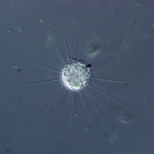
 Strahlengang bei DIC-Mikroskopie. Entscheidend sind die beiden Polarisationsfilter und die beiden Wollaston- oder Nomarski-Prismen.
Siehe auch
Weblinks
- Polarisations- und Interferenzkontrastmikroskopie, Website an der Uni Hamburg.
- Differential Interference Contrast, ausführliche Website der Firma Olympus mit interaktiven Applets (auf englisch).
Wikimedia Foundation.